Dr. Aruna Ashok MBBS, MS OG, DNB OG
- Clinical Director
 Aruna Ashok | 12 Mar 2022
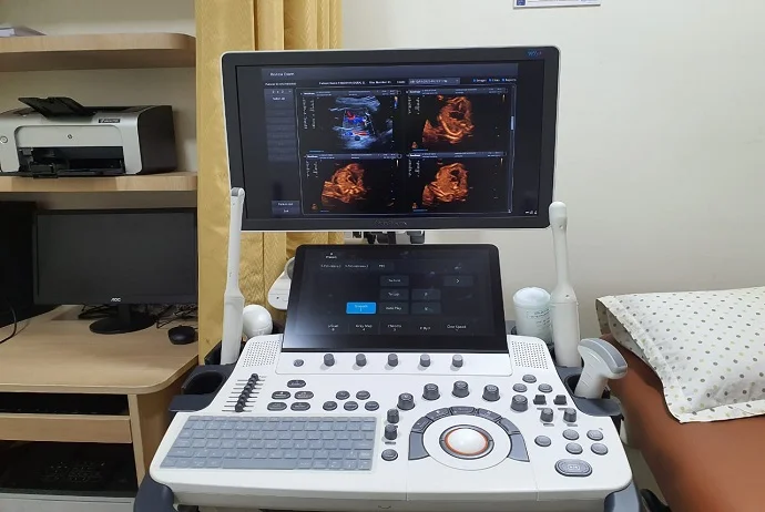
Ultrasound provides the image of the tissues, organs and the internal outlook of the body. It uses soundwaves to fulfill the various needs expected during scans and unlike X-ray, this does not use radiation. Ultrasound’s benefits are used in the many Ds that are part of our lives.
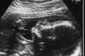
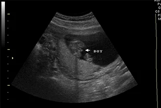
Ultrasound in this scenario shows the imagery of the baby in the womb. It shows the unborn baby’s position and development. It also checks for birth defects.
Here, ultrasound is used in checking the internal organ of the body and check for symptoms for diseases that haven’t yet surfaced like cancer.
It helps in early and clear detection of the symptoms for right diagnosis. Now let’s look at the many Ds in Ultrasound and their differences.
It gives a flat picturization with no depth. It is the most common ultrasound that people go to for having a look at their unborn baby. It gives a grey outline of the fetus and its organs.
The traditional ultrasound is also used in diagnosis like detecting heart disease. The scan might not be clear cut but it is helpful to some extent.
The imagery is a combination of many pictures of 2D ultrasound joined together to give an output like a photograph. This type of Ultrasound gives parents a clear image of the unborn baby’s face and not just the outline.
In the diagnostic hemisphere, it takes you to the next level and find things that escapes 2D Ultrasound. For instance, in infertility treatments, 3D pinpoints fibroids, polyps, or an adenomyosis.
Like other Ultrasounds, this also uses sound waves but 4D Ultrasound’s picturization is like a moving image. You can see the baby smile or yawn in the womb. And 5D Ultrasound brings high resolution into medicine.